As described in a recent post, dental conditions are often hidden and painful. The following posts are going to help describe conditions that pets can get as well as treatment. Many people I talk with are surprised how we can help pets with dental conditions and save teeth whenever possible.
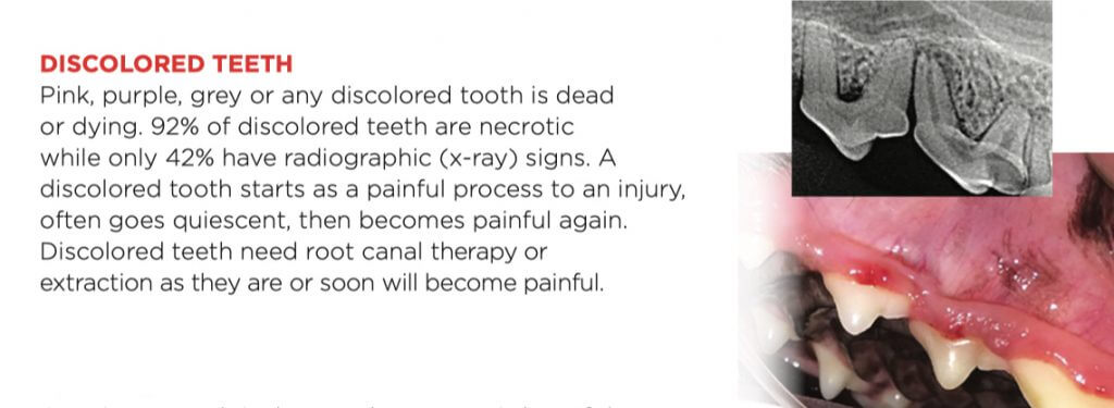
As alluded in the image text above, almost all discolored teeth are dead. They are discolored because the breakdown of the contents of the pulp leeches into the dentinal tubules discoloring the tooth. This is not just cosmetic as my father was taught, but a painful process. These breakdown products are a great place for bacteria to colonize exacerbating the problem. The only choices appropriate choices are root canal therapy or extraction.
As seen in the image above and its accompanying x-ray, teeth age like inverse trees. Instead of laying down rings on the outer edge of the trunk, dentin is laid down inside the tooth adding structure and decreasing the pulp (tooth center nerve and blood vessels) diameter. When a tooth stops laying down dentin and similar teeth nearby do not, one sees a wider pulp chamber in the dead tooth. Other signs can be bone loss related to disease at the tip of the root, root resorption, or narrowing of the pulp cavity due to calcification. One research paper states that only 42% of discolored teeth have x-ray signs, meaning that around 58% are still necrotic but don’t have x-ray signs yet.
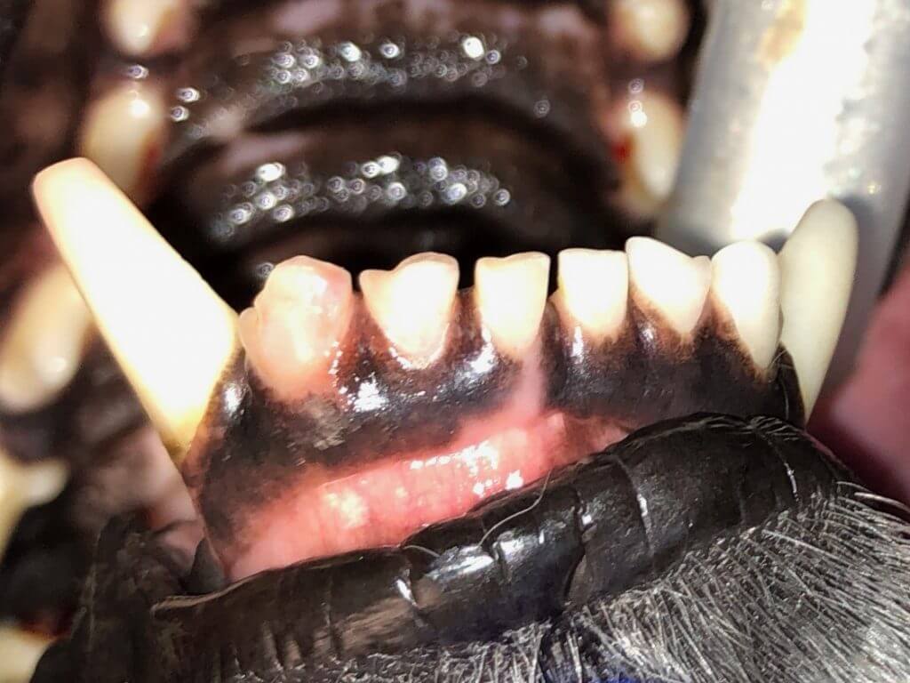
Another tool for evaluating a tooth to verify discoloration is called transillumination. It’s like candling an egg. A living tooth will have light shine through, while a dead one will not.
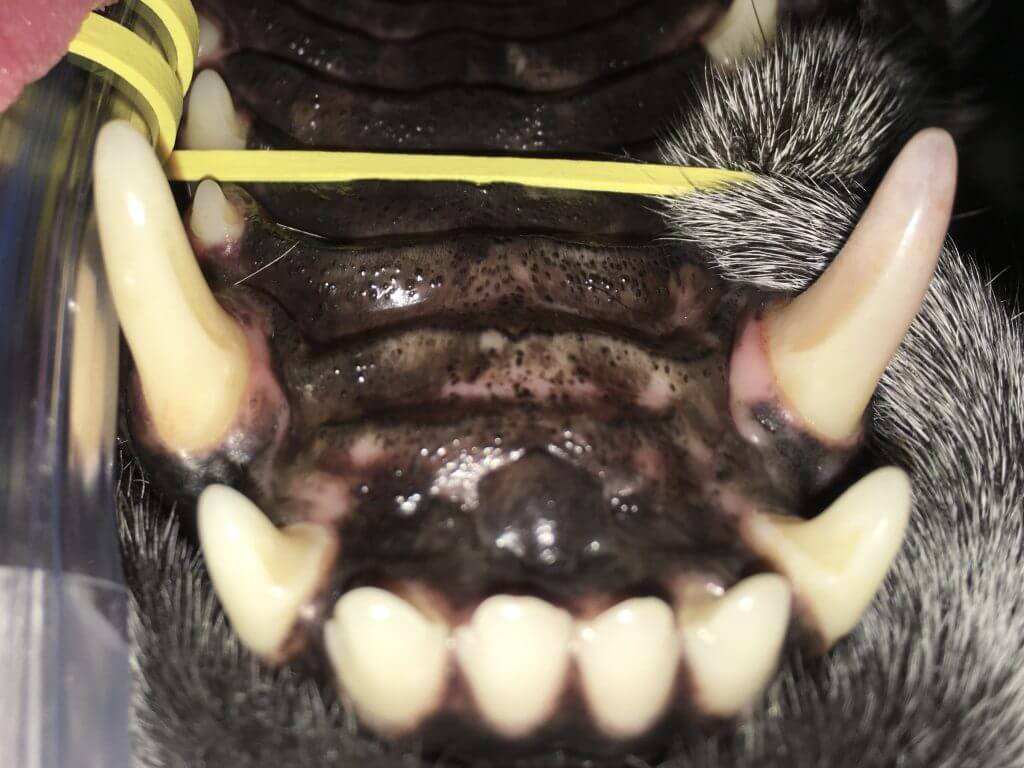
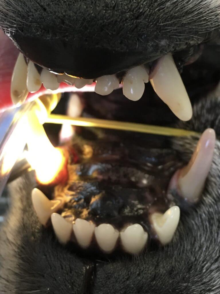
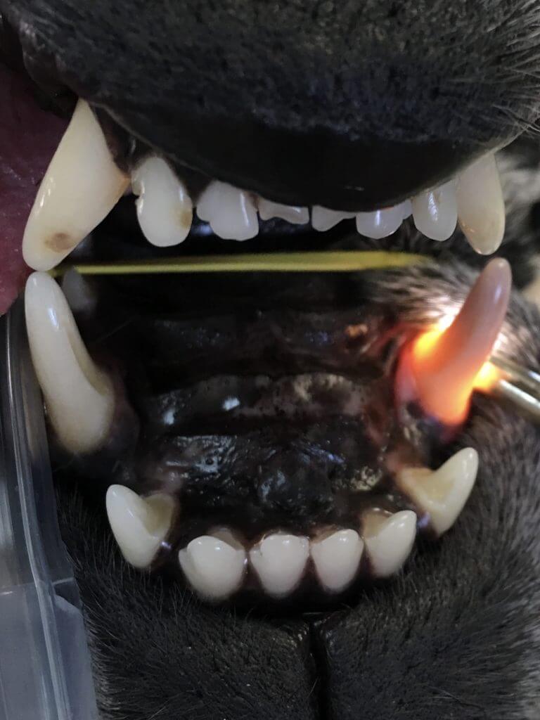
Although these images show obvious differences, it may not be this easy to tell. Transillumination alone cannot be used to make the judgment on treatment of a tooth. If one leaves behind a diseased tooth, the pet is left with pain or it will soon have pain at that site. There are systemic inflammatory effects (LINK to Increase Anes Safety on Advanced Dentistry page on fampetvet.com) known with diseases of the mouth, thus beyond pain, leaving behind disease is not wise. Extracting every affected tooth is preferable to leaving problems, but the most common teeth to become discolored are the canines and carnassials (the large important chewing teeth towards the back). There are a total of 8 of these structural teeth out of 42 total possible teeth in a dog’s mouth. The best course of action for the canines and carnassials is root canal therapy to save the structure and function but remove the pain and disease. One needs to be aware when deciding to extract these strategic teeth that they are losing almost all chewing function on that side of the mouth in the case of carnassials or removing a lot of jaw structure in the case of the canines. The most difficult tooth to extract in the entire mouth is a lower canine. This is one of the reasons that a common site for iatrogenic jaw fractures is adjacent to a lower canine extraction site. Root canal therapy must be considered, as it is best for the pet.
The exception to this discoloration discussion is reversible pulpitis. When a concussive force occurs to the body, usually a bruise is formed and resolves it. When a concussive force occurs to a tooth, if a pet were lucky enough to have a ‘bruise’ and still have the tooth continue to live, the breakdown products would still leech into the tubules discoloring the tooth. The veterinarian can prescribe antibiotics (amoxicillin-clavulanate or clindamycin) and non-steroidal anti-inflammatory drugs (provided the pet’s bloodwork is appropriate) for 7 days. If the tooth’s color doesn’t return to normal after two or three months, you can assume it’s irreversible discoloration (pulpitis) leaving the only appropriate choices of root canal therapy or extraction. If the tooth improves, it must be followed with intraoral x-rays in 4-6 months, then every 6-12 months. Continued discoloration is likely an irreversible process and 42% of the time x-rays will verify it. It does take 4-6 months for x-rays to show mineralization change, thus answers will not usually be known right away.
Contact us to learn more about root canal therapies in your pet.
